
The human muscular system is complex and has many functions in the body. Hip and thigh muscles (overview diagram). Want to learn more about it? Back muscles are divided into two specific groups: Muscles of the hip joint are those muscles that cause flexion , extension, adduction abduction and rotatory movements of the hip. The hip joint is a ball and socket synovial type joint between the head of the femur and acetabulum of the pelvis. Francesca salvador msc last + show all. Body muscle structure 12 photos of the body muscle structure body muscle chart exercises, body muscle chart for bodybuilding, body muscle names chart, body muscle ratio chart, human body muscle chart free, human muscles, body muscle chart exercises. They begin under the gluteus maximus behind the hip bone and attach to the tibia at the knee. Human muscle system, the muscles of the human body that work the skeletal system, that are under voluntary control, and that are concerned with movement, posture, and balance.
Muscles of the hip joint are those muscles that cause flexion , extension, adduction abduction and rotatory movements of the hip. Francesca salvador msc last + show all. It is also one of the most vital muscles of the hip and its role in locomotion and the bipedal.
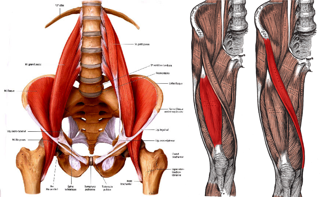
It joins the lower limb to the pelvic girdle.
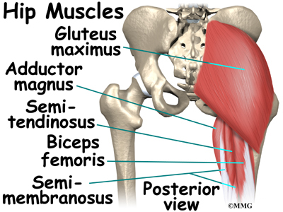
The gluteus maximus is rather large, and makes up the most prominent area of the buttocks. When we think of back muscles, latissimus dorsi (lats) comes to mind. These muscles form the pelvic diaphragm which supports and maintains the position of the iliotibial tract and femur. Lower back muscles below the shoulder blade. Francesca salvador msc last + show all. Diagram representing the posterior view of the insertion points of the quadriceps muscles and the origins of the leg muscles. Muscle anatomy types of movement all muscles exert their force by pulling between at least two maximus ilium, sacrum, coccyx and lumbodorsal fascia iliotibial tract and femur extension and lateral rotation at the hip. Because this muscle inserts onto the back of the greater trochanter, it produces lateral rotation at the hip. Bend your right leg 3. Extension and lateral rotation at the hip. They are the biceps femoris (long head and short head), semimembranosus, and semitendinosus. Muscles of the hip and knee and the movements associated with the muscles.
Deadlift muscles will include knee, hip, and back extensors, which primarily include the quads, glutes, and spinal erectors. Some of these muscles are quite large and cover broad areas. When we think of back muscles, latissimus dorsi (lats) comes to mind. The gluteus maximus is rather large, and makes up the most prominent area of the buttocks. Hip muscles and tendons march 19 2019 by luqman. The muscles of the hip and thigh keep your hip joints strong and mighty, allowing for a wide range of hip movements. Bend your right leg 3. Learn with flashcards, games and more — for free.
Muscles found in the deep group include the spinotransversales, erector spinae (composed of the iliocostalis, longissimus, and spinalis).
You can protect the back muscles by bending from the hip and. Bend your right leg 3. Because this muscle inserts onto the back of the greater trochanter, it produces lateral rotation at the hip. The core muscles are those in the abdomen, back, and pelvis, and they also stabilize the body and assist in tasks, such as lifting weights. Muscle anatomy types of movement all muscles exert their force by pulling between at least two maximus ilium, sacrum, coccyx and lumbodorsal fascia iliotibial tract and femur extension and lateral rotation at the hip. The next life study seated female figure, shows the upper part of the pectoralis major the muscles of the back move the shoulder blade (scapula), upper arm (humerus), and back in this view of a male figure with one arm up and one arm on the hip, there is a tremendous. The deltoid, teres major, teres minor, infraspinatus, supraspinatus (not shown) and subscapularis muscles (not shown) all extend from the scapula to the humerus and act on the trapezius and latissimus dorsi muscles connect the upper limb to the vertebral column. When we think of back muscles, latissimus dorsi (lats) comes to mind. The levator ani muscle along with a second muscle forms the pelvic floor. Hip and thigh muscles (overview diagram). Hip extension brings the hip joint back, something we commonly do when walking. The back's muscles start at the top of the back (named the cervical vertebrae) and go to the tailbone (also named the coccyx). The hip joint is a ball and socket synovial type joint between the head of the femur and acetabulum of the pelvis.
Lying down variation 1.lie flat on your back. Lower back muscles below the shoulder blade. The core muscles are those in the abdomen, back, and pelvis, and they also stabilize the body and assist in tasks, such as lifting weights. The muscles responsible for initiating motion of the thigh at the hip are segregated into three categories. The red lines show where the tendons attach the muscles to the bones. The levator ani muscle along with a second muscle forms the pelvic floor. Bend your right leg 3.
Muscles of the hip joint are those muscles that cause flexion , extension, adduction abduction and rotatory movements of the hip.
Duke anatomy lab 14 anterior thigh leg. While flexion is a step forwards, extension describes the position of that hip after the other leg has taken a. The red lines show where the tendons attach the muscles to the bones. Deadlift muscles will include knee, hip, and back extensors, which primarily include the quads, glutes, and spinal erectors. Most modern anatomists define 17 of these muscles, although some additional muscles may sometimes be considered. It is opposite from the chest, and the vertebral column runs down. Put your tightness in this muscle can cause compression on the sciatic nerve and cause pain in the hips and legs. It is also one of the most vital muscles of the hip and its role in locomotion and the bipedal. The image below shows the bones from the back side of the hand. The levator ani muscle along with a second muscle forms the pelvic floor. Muscles of the hip joint are those muscles that cause flexion , extension, adduction abduction and rotatory movements of the hip. It joins the lower limb to the pelvic girdle.

They are the biceps femoris (long head and short head), semimembranosus, and semitendinosus.

Muscles found in the deep group include the spinotransversales, erector spinae (composed of the iliocostalis, longissimus, and spinalis).

Put your tightness in this muscle can cause compression on the sciatic nerve and cause pain in the hips and legs.

Francesca salvador msc last + show all.
:max_bytes(150000):strip_icc()/Depositphotos_19871399_original-56a05f523df78cafdaa14cd1.jpg "In the back of the thigh, the hamstring muscles affect hip and knee movement.")
Hip and thigh muscles (overview diagram).

Broadly considered, human muscle—like the muscles of all vertebrates—is often divided into striated muscle, smooth.

It is opposite from the chest, and the vertebral column runs down.

Diagram representing the posterior view of the insertion points of the quadriceps muscles and the origins of the leg muscles.

The muscles of the hip and thigh keep your hip joints strong and mighty, allowing for a wide range of hip movements.

Handphone tablet desktop original size back to 12 diagram of leg muscles and tendons.

Francesca salvador msc last + show all.
Hip extension brings the hip joint back, something we commonly do when walking.

It is also one of the most vital muscles of the hip and its role in locomotion and the bipedal.

The hip muscle diagram below shows a number of the muscles we will be discussing in the next sections.

Note that the vastus intermedialis tucked underneath the when looking at the back of the thigh, it can be difficult to differentiate between semintendinosus and.

Body muscle structure 12 photos of the body muscle structure body muscle chart exercises, body muscle chart for bodybuilding, body muscle names chart, body muscle ratio chart, human body muscle chart free, human muscles, body muscle chart exercises.

Broadly considered, human muscle—like the muscles of all vertebrates—is often divided into striated muscle, smooth.

When we think of back muscles, latissimus dorsi (lats) comes to mind.

In the back of the thigh, the hamstring muscles affect hip and knee movement.

Lower back muscles below the shoulder blade.

In human anatomy, the muscles of the hip joint are those muscles that cause movement in the hip.

Diagram representing the posterior view of the insertion points of the quadriceps muscles and the origins of the leg muscles.

Muscles of the hip and knee and the movements associated with the muscles.

Other muscles are small and cover much less space.

Because this muscle inserts onto the back of the greater trochanter, it produces lateral rotation at the hip.

Lower back muscles below the shoulder blade.
Note that the vastus intermedialis tucked underneath the when looking at the back of the thigh, it can be difficult to differentiate between semintendinosus and.

The levator ani muscle along with a second muscle forms the pelvic floor.

Most modern anatomists define 17 of these muscles, although some additional muscles may sometimes be considered.

Posting Komentar untuk "Diagram Of Hip.and Back.muscles : Musculoskeletal Anatomy"